X-Ray & Lab Investigations for Accurate Orthopaedic Diagnosis in Mumbai
Precise diagnosis is the foundation of effective orthopaedic treatment. Dr. Bafna's clinic in Mumbai utilises advanced imaging and laboratory investigations — from digital X-rays and MRI scans to comprehensive blood panels — to identify the exact cause of your bone, joint, or muscle condition before any treatment begins.
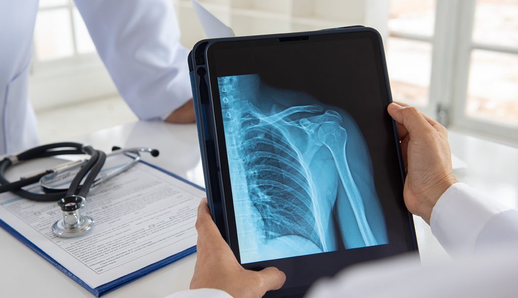
Digital X-Ray for Orthopaedic Diagnosis
X-ray imaging remains the first and most essential diagnostic tool in orthopaedic practice. At Dr. Bafna's clinic in Borivali, Mumbai, digital X-rays are used to evaluate the structural integrity of bones and joints — providing rapid, high-resolution images that reveal fractures, dislocations, joint space narrowing, bone deformities, and signs of degenerative arthritis with exceptional clarity.
- Detects fractures, hairline cracks, stress fractures, and bone displacements in all major joints and long bones.
- Evaluates joint space to identify early and advanced stages of osteoarthritis, rheumatoid arthritis, and cartilage loss.
- Assesses post-operative implant positioning, bone healing progress, and hardware integrity after orthopaedic surgery.
Key Benefits
- Delivers immediate, high-definition bone images that guide accurate diagnosis within minutes of the scan.
- Weight-bearing X-rays assess how joints behave under normal load — critical for knee and hip arthritis grading.
- Digital format allows instant report sharing, precise measurements, and long-term comparison across follow-up visits.
Procedure
The X-ray procedure is quick, painless, and requires no preparation. Patients are positioned to capture the specific joint or bone region of concern in one or more planes. Dr. Bafna personally reviews and interprets all imaging in the context of the patient's clinical examination, ensuring that findings are accurately correlated with symptoms to guide the most appropriate treatment plan in Mumbai.
Frequently Asked Questions
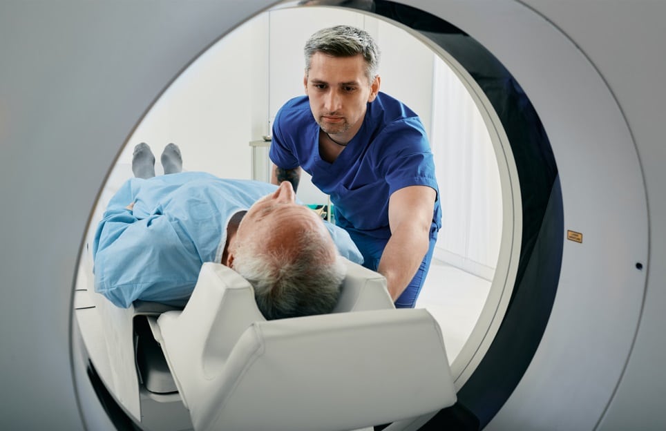
MRI & CT Scan for Orthopaedic Conditions
While X-rays excel at visualising bone, MRI and CT scans provide critical detail about soft tissues, cartilage, spinal structures, and complex fracture patterns that plain X-rays cannot show. Dr. Bafna routinely advises MRI or CT investigations in Mumbai when a deeper understanding of joint pathology, nerve compression, ligament tears, or pre-surgical anatomy is required for precise orthopaedic planning in Borivali, Mumbai.
- MRI provides detailed imaging of ligaments, tendons, cartilage, intervertebral discs, and bone marrow — ideal for knee, shoulder, spine, and hip conditions.
- CT scan delivers precise cross-sectional bone anatomy, essential for complex fracture assessment, joint reconstruction planning, and spinal surgery preparation.
- CT angiography evaluates blood vessel integrity around major injuries, critical for complex trauma management and limb-salvage surgery.
Key Benefits
- Identifies soft tissue injuries — ACL tears, rotator cuff ruptures, meniscal damage — that are completely invisible on X-ray.
- 3D CT reconstructions allow surgeons to plan implant sizing, screw trajectories, and bone cuts with high precision before entering the operating theatre.
- MRI detects early avascular necrosis, bone marrow oedema, and stress reactions before they become visible on X-ray — enabling earlier, less invasive intervention.
Procedure
MRI and CT scans are non-invasive and typically completed within 20–45 minutes. Dr. Bafna advises the most appropriate scan based on the clinical diagnosis and prescribes specific imaging protocols — such as contrast enhancement or dedicated joint sequences — to ensure that the resulting images provide maximum diagnostic value for each individual patient in Mumbai.
Frequently Asked Questions
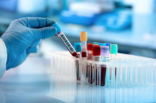
Laboratory & Blood Investigations
Laboratory and blood investigations are an integral part of comprehensive orthopaedic diagnosis. Many bone and joint conditions — including inflammatory arthritis, infections, metabolic bone disease, and nutritional deficiencies — manifest through specific abnormalities in blood markers. Dr. Bafna's clinic in Mumbai recommends targeted lab investigations to confirm diagnoses, guide medication choices, and monitor treatment response over time in Borivali, Mumbai.
- Inflammatory markers — ESR, CRP, and RA factor — to diagnose and monitor rheumatoid arthritis, gout, and infectious joint conditions.
- Serum calcium, phosphorus, alkaline phosphatase, and Vitamin D levels to evaluate metabolic bone diseases such as osteoporosis and rickets.
- Pre-operative blood panels — CBC, coagulation profile, blood group, and renal function — to ensure patient safety before any elective orthopaedic surgery.
Key Benefits
- Identifies systemic causes of joint pain — such as gout, lupus, or septic arthritis — that require medical rather than surgical management.
- Monitors response to disease-modifying drugs and anti-inflammatory medications, enabling dose adjustments for optimal therapeutic effect.
- Detects Vitamin D and calcium deficiencies early, allowing supplementation that protects bone density and reduces fracture risk significantly.
Procedure
Blood tests are prescribed based on Dr. Bafna's clinical assessment of each patient in Mumbai. A small venous blood sample is collected under aseptic conditions and processed at a certified diagnostic laboratory. Results are reviewed by Dr. Bafna in conjunction with imaging findings and clinical examination to formulate a complete, evidence-based orthopaedic diagnosis and treatment plan.
Frequently Asked Questions
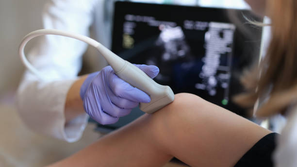
Bone Density Scan & Musculoskeletal Ultrasound
Bone density scanning (DEXA) and musculoskeletal ultrasound are two highly valuable diagnostic tools that complement X-ray and MRI in orthopaedic practice. Dr. Bafna recommends these investigations in Mumbai for patients at risk of osteoporosis, those with tendon or soft tissue injuries, and individuals requiring real-time dynamic assessment of joint structures during movement in Borivali, Mumbai.
- DEXA scan accurately measures bone mineral density at the hip and spine, producing a T-score that classifies bone health as normal, osteopenic, or osteoporotic.
- Musculoskeletal ultrasound provides real-time imaging of tendons, ligaments, bursae, and muscle tissue — ideal for diagnosing rotator cuff tears, plantar fasciitis, and tendinopathies.
- Ultrasound-guided procedures — including diagnostic joint aspirations and therapeutic injections — are performed with precision using live imaging for maximum accuracy.
Key Benefits
- DEXA scanning identifies bone loss years before a fragility fracture occurs, enabling preventive treatment that dramatically reduces future fracture risk.
- Ultrasound involves no radiation, making it ideal for repeated assessments and for use in children, pregnant women, and elderly patients.
- Dynamic ultrasound assessment captures how tendons and joints behave during movement — information that static imaging modalities cannot provide.
Procedure
A DEXA scan is a brief, comfortable procedure taking 10–20 minutes in which the patient lies still while low-dose X-ray beams measure bone density at the spine and hip. Musculoskeletal ultrasound is performed in clinic using a handheld probe applied with gel to the skin over the area of concern. Both investigations are painless and provide immediate results that Dr. Bafna correlates with clinical findings to plan the most effective orthopaedic intervention in Mumbai.